
LASER and IPL TREATMENTS
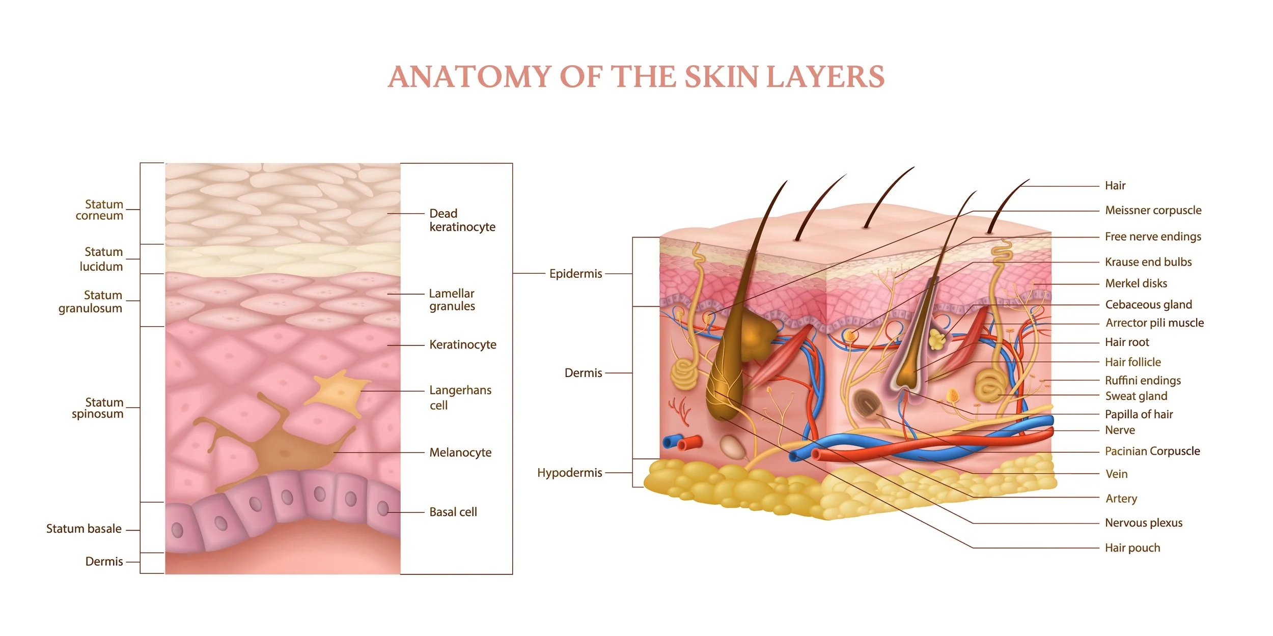
Anatomy of the Skin
The Epidermis
The epidermis is where skin cells are born and pushed through their natural cycle to where they keratinize and shed. The deepest level of the epidermis is known as the stratum basale and cells grow here because they are close to their nutrient source(in the dermis), as they are pushed up and away they begin to die(the stratum granulosum). After they die they start to fill up with Keratin.
Merkel Cells
Merkel Cells have a function in sensory pain and pressure. They are special skin cells that help you feel touch. They're like tiny sensors in your skin, especially in areas like your fingertips. When something presses against your skin, Merkel cells send signals to your brain so you can feel textures, shapes, and pressure. They are found in the stratum basale, where skin cells are born.
As you grow older, the number of merkel cells decrease. This is important to take into account when working on younger kids and adults, they will likely feel the laser more.
Langerhans Cells
Langerhans cells are dendritic cells that are responsible for the immunity of your skin. They are susceptible to heat, meaning they will move away from a heat source for up to a week. This is important as its important to advise your client to continue cooling the treated area after leaving the clinic/spa to allow for a better and faster recovery.
Skin Anatomy
There are 3 layers to skin:
Epidermis
Thinnest layer of the skin with a depth of 0-150 microns (this is an estimate and varies from patient to patient)
Indications: Tone, texture improvements
Strongest layer of the skin
doesn’t have vascularization, therefore it relies on absorbing oxygen and nutrients from the blood vessels in the connective tissue.
Dermis
Depth of 150-700 microns (varies patient to patient)
Indications: Fine Lines
Most of the body’s collagen is found in this layer
Stretch marks are caused by tears in the collagen of this layer
Hypodermis (Subcutaneous Layer)
Depth 700 microns and beyond
The “structure” of the skin
Home of dormant fibroblasts and melanocytes
Indications: Deeper wrinkles, scars.
Conditions of the Skin
Blisters
When you rub the skin, it stimulates cell division = callous
If you rub it too hard, the stratum basale tears away from the basement membrane and causes a gap, which is filled with fluid.
The epidermis then dies because it’s too far away from nutrients.
Remember the epidermis has no vascularization
Smaller blisters, less than 5 mm in diameter are knows as vesicles
Larger blisters, greater than 5 mm in diameter are know as bullae
Skin Cancer
Skin cancer is the most common cancer in the USA. The major risk facto of all skin cancers is ultraviolet light.
Three major ypes:
1) Basal Cell Carcinoma
Cancer of the blood vessels in the dermis
the most common type of skin cancer
accounting to about 75% of skin cancers
Almost never metastasizes or crosses the basement membrane, so almost never fatal
The most easily cured through surgical removal, no chemotherapy or radiation is usually needed
Symptoms: shiny nodules, usually on the nose or other sun exposed areas
2) Squamous Cell Carcinoma
about 25% of all skin cnacers
Will metastasize if not treated
Actinic Keratosis
This is a premalignant condition of thick, scaly or crusy pathes of skin
it’s more common in fair skin, in sun-exposed areas
20% of these lesions progress to squamous cell carcinoma
Begin as flat, dry, scaly areas and later grow into tough, wart like lesions
We DO NOT want to stimulate these with heat. Always ask for a doctors note if you are unsure of any skin conditions
3) Melanoma
Cancer of the melanocytes of the epidermis
about 1% of skin cancer
Highly metastatic, causes 75% of skin caner deaths
Asymmetrical, sharp but irregular borders and edges
Not uniform in colour
ABCD’s of Skin Cancer
A - Asymmetry, where one side of the mole looks different than the other side
B - Border, where the border is jagged or irregular instead of smooth and regular
C - Color, where the mole contains more than one colour
D - Diameter, a mole that’s smaller than the eraser on the end of a pencil (6mm) generally is not a cause for concern. However, a larger mole is not necessarily a dangerous one, a larger mole can grow to be bigger than 6mm. Look for the other signs as well
E - Elevated, is not flat nut lifted.
F - Feels, has texture to it, not uniform or “soft”
Eczema
Itchy red skin that comes and goes. It’s caused by an autoimmune reaction.
The most frequently occurring for of eczema is Atopic Dermatitis.